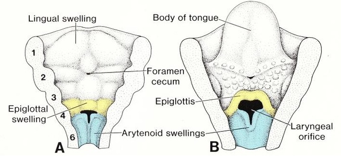
Figure 2: The larynx is develops from the 4th and 6th pharyngeal. Available at archeshttp://www.ultratwistersgym.com/Resources/Respiratory/Respiratory.html
The endodermal tissues of the cranial end of the laryngotracheal tube made up the epitheal lining of the larynx.
The larynx has cartilages surrounding it and it is develop from the mesenchyme of the fourth and the sixth pairs of pharyngeal arches (A,B). It is from the neural crest cells.
Arytenoid swelling is formed at the cranial end of the laryngotracheal tube by the proliferation of the mesenchymal tissue (derived from neural crest cells). It will grow towards the tongue and forms the primordial glottis. As it grows further, it changes the primordial glottis into a T-shaped laryngeal inlet.
Temporary occlusion of the laryngeal inlet occurs due to rapid proliferation of the epithelial lining of the larynx. By the 10th week of gestation, recanalization occurs and consequently, pair of laryngeal ventricles are formed.
The laryngeal ventricles are bound by tissue that progress into false and true vocal cords.
Proliferation of the mesenchyme in the ventral part of the 3rd and 4th pharyngeal arches will formed hypopharyngeal eminence with later form the epiglottis.
The superior laryngeal nerve will supplies the part of larynx that develop from the 4th pharyngeal arches. The recurrent laryngeal nerve will supplies the part of larynx that derives from the 6th pharyngeal arches. The 2 nerves are branches of the vagus nerve (CN X).
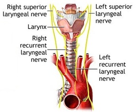
Figure 3: Nerve supplies of the larynx is from CN X (Vagus nerve). Available at http://svpow.com/2011/05/23/the-worlds-longest-cells-speculations-on-the-nervous-systems-of-sauropods/
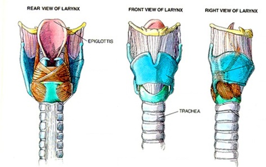
Figure 4: Adults larynx. Available at http://www.edoctoronline.com/medical-atlas.asp?c=4&id=21680